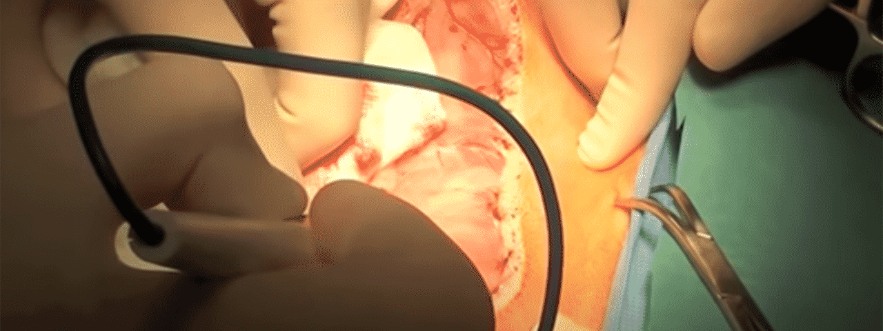
We feature a range of vet surgical courses depending on the areas a vet surgeon wants to specialize in.
Dr Charles Kuntz, Specialist Surgeon for VetDojo explains the procedure on how to remove fibrosarcoma from the maxilla of a dog.
In this operation, Dr Kuntz also performed a temporary carotid artery ligation to prevent haemorrhage during surgery.
Fibrosarcoma is one of the common tumours we see in the mouth of dogs. It is associated with a high risk of local recurrence and about 25% chance of metastasis (spread) to the lungs.
How to perform carotid ligation and maxillectomy on a dog
A ventral midline cervical approach is made to expose the carotid arteries.
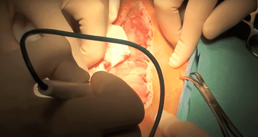
The sternohyoideus and sternothyroideus muscles are separated.
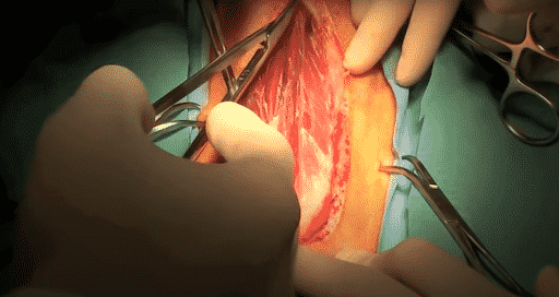
A large Gelpi Retractor is used to maintain retraction of the muscles.
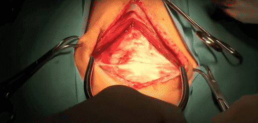
The carotid artery is isolated using right angle forceps and exteriorised.
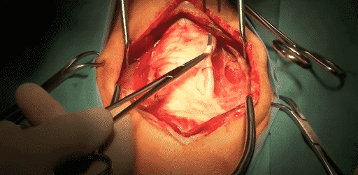
A piece of O-silk is passed around the carotid artery. The same procedure is repeated on the opposite side.
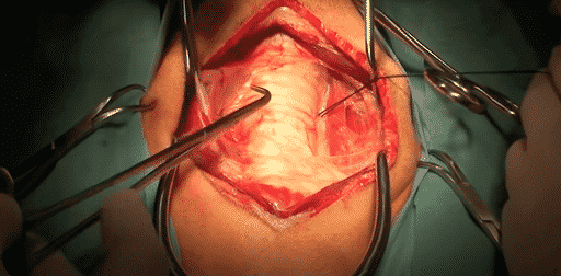
A Rommel tourniquet is created by passing a piece of eight to ten French urinary catheter through the skin. The same procedure is repeated on the opposite side.
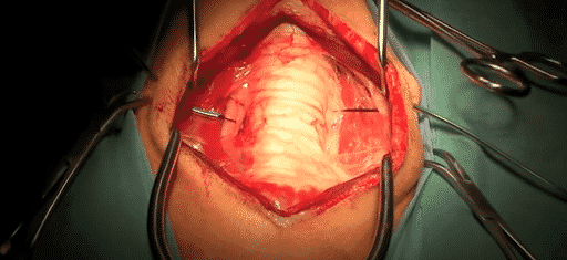
The incision is closed in routine fashion.
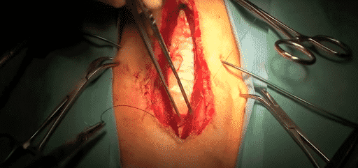
Just before starting the maxillectomy, the Rommel tourniquets are pulled tight and affixed in place using the hemostats.
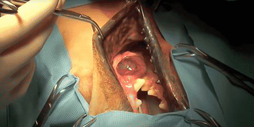
Position of the patient has changed and a cheiloplasty is performed to explore the maxilla.
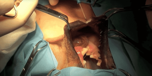
The freely movable gingiva is separated from the maxilla to facilitate closure of the tumour. Great care is taken to avoid contaminating surgical margins with tumour cells.
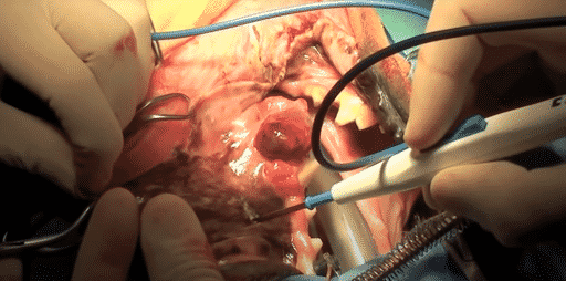
Large maxillary vessels should be identified and ligated prior to transection.
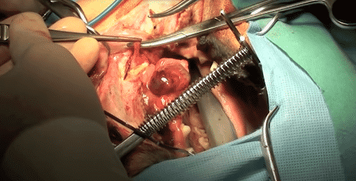
This area of the zygomatic arch is isolated and an osteotomy is performed using an oscillating saw. The osteotomy is continued using the oscillating saw at a distance of three centimetres away from any visible tumour.
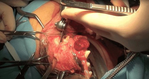
The osteotomy of the palatine bone is performed using a very sharp osteotome and a mallet.
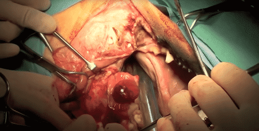
The piece of bone is then separated away from the maxilla and haemorrhage is controlled using ligation.

The buccal mucosa is sutured to the palatine mucosa using cruciate sutures of 2-0 PDS. The upper and lower buccal mucosa is sutured using 2-0 PDS in a simple continuous pattern.
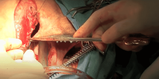
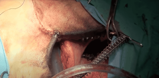
Routine subcutaneous and intradermal sutures are then completed.
Vetdojo is a new e-learning platform which provides academic information distilled into the real world, practical skills that veterinarians can apply in a clinical setting.
Students have access to informative and educational surgical content, mentorship, and support plus ability to connect with a community of like-minded vets to share challenges and questions. To view the range of courses or start one today visit www.vetdojo.com.

